Understanding the 4 Types of Kidney Stone Surgery: Which One is Right for You?
Kidney stones are a common medical condition affecting millions of people worldwide. They can cause severe pain and discomfort, and if left untreated, can lead to serious urinary tract complications or kidney damage. While small stones can often pass naturally with hydration and medication, larger or obstructive stones frequently require surgical intervention.
Here is a comprehensive breakdown of the four primary types of kidney stone surgery, how they work, their advantages, and when each is recommended.
1. Extracorporeal Shock Wave Lithotripsy (ESWL)
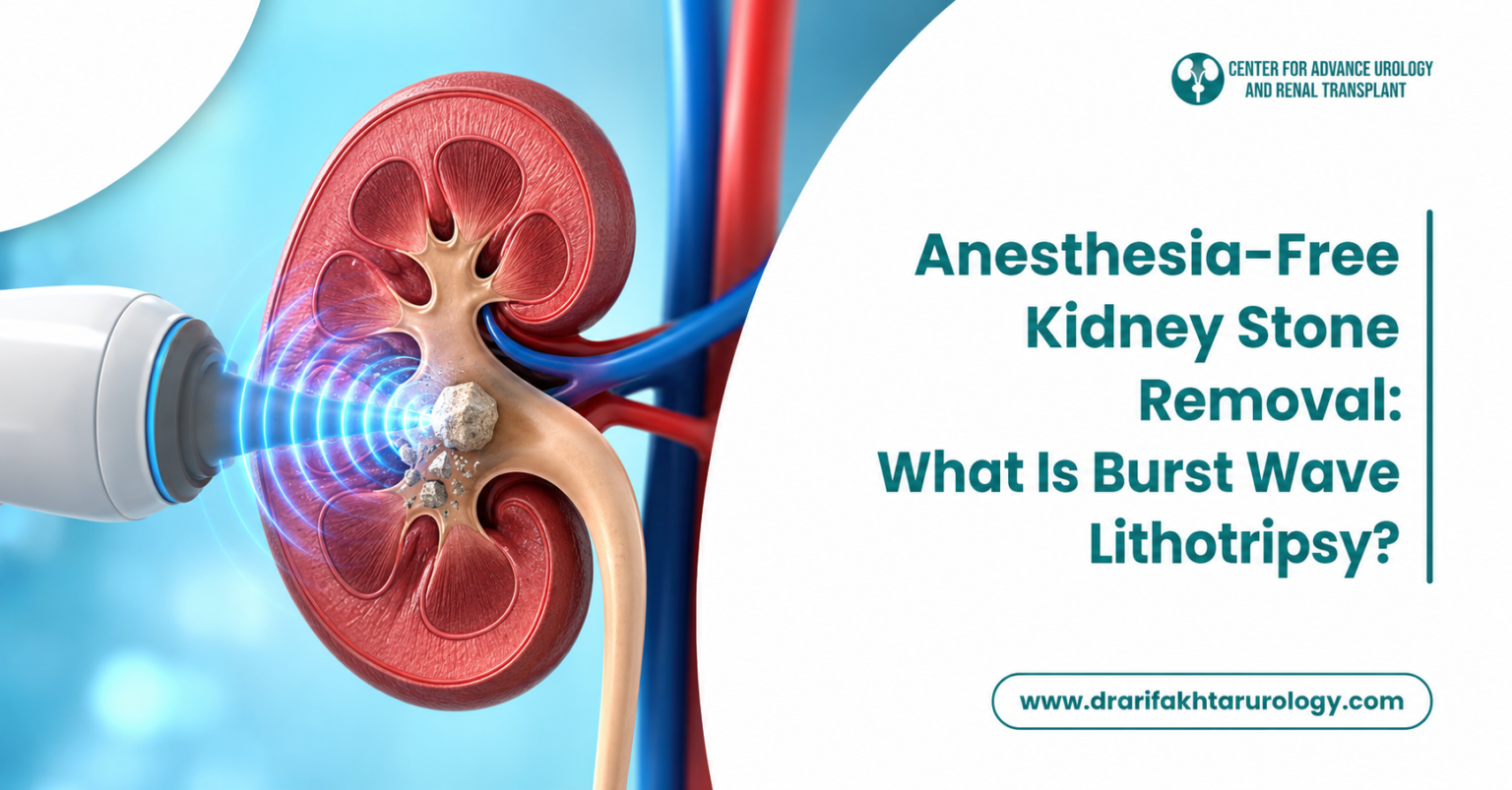
Extracorporeal Shock Wave Lithotripsy (ESWL) is a non-invasive procedure commonly used to treat kidney stones and certain ureteral stones. Because it requires no incisions or internal scopes, it is often the first-line surgical choice for eligible patients.
How It Works
Preparation: The patient receives a light sedative or anesthesia to remain comfortable and still during the procedure.
Targeting: The patient lies on a water-cushioned table. The surgeon uses real-time X-ray (fluoroscopy) or ultrasound to pinpoint the exact location of the stone.
Fragmentation: The lithotripter delivers focused, high-energy acoustic shock waves through the skin directly to the stone, breaking it down into tiny, sand-like fragments.
Passage: Over the following days to weeks, these small fragments pass naturally out of the body through the urine.
Advantages
- Non-Invasive: No surgical cuts, punctures, or internal scopes are required.
- Outpatient Procedure: Most patients return home the same day.
- High Efficacy for Moderate Stones: Highly effective for small-to-medium stones (typically under 20 mm in diameter) located in the kidney or upper ureter.
Risks and Limitations
Not ideal for very hard stones (e.g., cystine or calcium oxalate monohydrate) or stones larger than 20 mm.
Passing the broken fragments through the ureter can cause temporary pain or mild hematuria (blood in urine).
Some patients may require multiple sessions or a temporary ureteral stent to facilitate drainage.
2. Percutaneous Nephrolithotomy (PCNL)
Percutaneous Nephrolithotomy (PCNL) is a minimally invasive surgical procedure designed specifically for large, dense, or complex kidney stones (such as staghorn calculi).
How It Works
Anesthesia: The procedure is performed in an operating room under general anesthesia, typically with the patient lying face down (prone).
Access Creation: Using fluoroscopic or ultrasound guidance, the surgeon inserts a needle through a small incision in the patient’s back directly into the kidney’s collecting system.
Tract Dilation: A guidewire is placed, and dilators create a narrow channel (tract) into the kidney.
Stone Extraction: A specialized scope called a nephroscope is inserted through the tract. Larger stones are broken up using ultrasonic, pneumatic, or laser energy, and the fragments are suctioned or grasped and removed entirely.
Advantages
Gold Standard for Large Stones: The most effective method for stones larger than 2 cm (20 mm) or irregularly shaped stones.
Direct Removal: Unlike ESWL, the fragments are extracted during the procedure rather than left to pass through the urinary tract.
High Clearance Rate: Offers the highest stone-free rate in a single setting for massive stones.
Risks and Limitations
Requires an overnight hospital stay (typically 1 to 2 days).
Higher risk of post-operative bleeding or infection compared to non-invasive options.
Potential need for a temporary nephrostomy tube or internal stent to drain the kidney while it heals.
3. Ureteroscopy (URS / RIRS)
Ureteroscopy is an endoscopic procedure used to diagnose and remove stones located anywhere along the urinary tract, particularly in the ureter or lower kidney poles.
How It Works
Access via Natural Pathways: While the patient is under general or spinal anesthesia, a long, thin, flexible scope (ureteroscope) is guided through the urethra, bladder, and up into the ureter or kidney.
Visualization: A miniature camera on the tip of the scope provides real-time video of the urinary tract and stone.
Fragmentation and Retrieval: Small stones are captured intact using a miniature wire basket. Larger stones are pulverized into fine dust using an advanced holmium or thulium laser fiber.
Stent Placement: A temporary plastic tube (double-J stent) is often placed in the ureter for a few days to prevent swelling and ensure proper urine flow.
Advantages
No Incisions: Utilizes the body's natural urinary pathways.
Versatile: Can treat stones throughout the entire ureter as well as inside the kidney (Retrograde Intrarenal Surgery - RIRS).
Same-Day Discharge: Usually performed as an outpatient procedure.
Risks and Limitations
Temporary bladder irritation, mild cramping, or blood in the urine while the stent is in place.
Rare risks include ureteral perforation, stricture formation, or urinary tract infection.
4. Open Kidney Stone Surgery
Open kidney stone surgery (open nephrolithotomy or pyelolithotomy) involves accessing the kidney or ureter through a traditional surgical incision. With modern advancements in endourology, open surgery is rarely required and represents less than 1–2% of all stone cases.
When Is It Considered?
Anatomical Complexities: Severe kidney malformations or spinal deformities that make endoscopic access impossible.
Extremely Large/Complicated Stones: Giant staghorn stones that cannot be safely managed with multiple PCNL procedures.
Concurrent Conditions: When kidney stone removal is combined with another open corrective surgery (e.g., repair of ureteropelvic junction obstruction).
Failed Prior Procedures: Cases where minimally invasive approaches have repeatedly failed.
Risks and Recovery
Requires general anesthesia and a large flank or abdominal incision.
Longer hospital stay (typically 3 to 7 days) and an extended recovery period of 4 to 6 weeks.
Increased postoperative pain, risk of wound infection, and incisional hernia.
Quick Comparison Guide
| Procedure | Invasiveness | Best For | Typical Recovery Time | Anesthesia |
| ESWL | Non-invasive (External waves) | Small to medium stones (<2 cm) | 1–3 days | Sedation / Light |
| PCNL | Minimally invasive (Small back incision) | Large (>2 cm) or staghorn stones | 1–2 weeks | General |
| Ureteroscopy | Minimally invasive (Natural openings) | Mid-to-lower ureteral and kidney stones | 2–5 days | General / Spinal |
| Open Surgery | Traditional open surgical incision | Highly complex or anatomically abnormal cases | 4–6 weeks | General |
Frequently Asked Questions
Which kidney stone surgery has the fastest recovery time?
ESWL and Ureteroscopy offer the shortest recovery windows. Most patients return to normal daily routines within 2 to 4 days, whereas PCNL requires about a week or two, and open surgery requires several weeks.
How do doctors decide which procedure is right for me?
Urologists base their recommendation on four key factors:
Stone size and number
Stone location (upper kidney, lower kidney, or ureter)
Stone density/composition (visible on a non-contrast CT scan)
Your general health and urinary tract anatomy
Will I need a stent after kidney stone surgery?
A temporary ureteral stent (DJ stent) is frequently placed after ureteroscopy and occasionally after PCNL or ESWL. It prevents the ureter from swelling shut and helps residual stone dust pass without causing severe renal colic. It is usually removed in the clinic within 3 to 10 days.
Final Thoughts
Kidney stone surgery has evolved into a predominantly minimally invasive field, offering high success rates with minimal discomfort. The right procedure depends entirely on the unique size, density, and location of the stone. Consult a qualified urologist to review your diagnostic imaging and discuss the safest, most effective path forward. Once treated, prioritizing long-term prevention—primarily consistent hydration and dietary adjustments—remains the most critical step in avoiding future stones.