
Dr. Arif Akhtar
Senior Urologist
Hello! 👋 Welcome to Dr. Arif Akhtar. He is a Gurugram-based urologist specializing in robotic urology, uro-oncology, and kidney transplantation.
Just now
Kidney stones are one of the most common reasons patients see a urologist — and one of the most treatable, once the stone's size, location and composition are properly assessed. Dr. Arif Akhtar treats kidney stones in Gurugram using the full range of modern options, from monitoring small stones to minimally invasive procedures like RIRS and PCNL for larger or complex stones. This page explains what kidney stones are, how they're diagnosed, the treatment options available, and what to expect if you need a procedure.
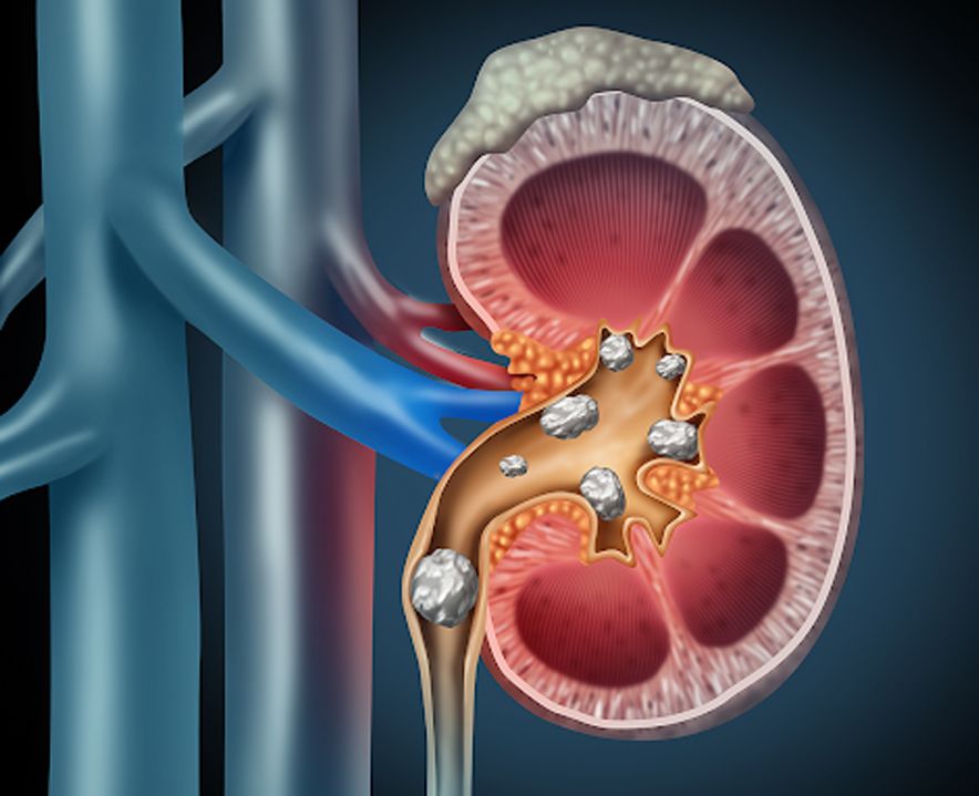
Kidney stones (renal calculi) are hard deposits that form when minerals and salts in the urine crystallise inside the kidney. The most common type is calcium oxalate; less common types include uric acid stones, struvite stones (often linked to infection) and cystine stones (an inherited condition). Knowing the stone type matters because it affects both treatment choice and how future stones can be prevented.
Schedule a Clinical EvaluationKidney stones can cause sudden and severe symptoms, although some stones may not cause any symptoms at all. Common symptoms include:
Sudden, severe pain in the flank or lower back, often radiating to the groin
Blood in the urine (visible or detected on a urine test)
Pain or burning while urinating
Nausea or vomiting during an acute episode
Frequent urge to urinate, or urine that looks cloudy or unusually coloured
In some cases, no symptoms at all — the stone is found incidentally on a scan
Understanding Factors Associated with Kidney Stones
Kidney stones can develop due to a combination of hydration, dietary, metabolic, lifestyle and genetic factors. Understanding these risk factors can help with appropriate treatment and strategies to reduce the chance of future stones.
Chronic dehydration can make urine more concentrated, increasing the likelihood of crystal formation and kidney stones.
A diet high in oxalate, sodium or animal protein may increase the risk of developing certain types of kidney stones.
A personal or family history of kidney stones can increase the likelihood of developing stones again.
Certain metabolic conditions and urinary tract infections can contribute to the formation of kidney stones.
Obesity and low levels of physical activity may be associated with a higher risk of kidney stone formation.
Some medications and dietary supplements may affect urine composition and contribute to kidney stone formation.
Diagnosis typically starts with a urine test and blood tests to check kidney function, followed by imaging — usually an ultrasound or a non-contrast CT scan (CT KUB) — to confirm the stone's size, exact location and number. In select cases, further metabolic work-up helps identify why stones are forming, particularly for patients with recurrent stones.
Many small stones (generally under about 5 mm) can pass on their own with hydration and pain management, monitored by your urologist. Larger stones, stones causing persistent pain or blockage, stones associated with infection, or stones that aren't moving on their own usually need an active procedure. The right approach depends on stone size, exact position in the kidney or ureter, and your overall health.
Kidney stone treatment depends on the stone's size, location, composition, kidney anatomy and your overall health. Dr. Arif Akhtar evaluates your imaging and symptoms to recommend the most appropriate approach, ranging from watchful waiting and medical management to minimally invasive procedures.
For small stones likely to pass naturally, treatment may simply involve fluids, pain relief and medication to help the stone move, with follow-up imaging to confirm it has passed.
Extracorporeal shockwave lithotripsy uses focused sound waves from outside the body to break a stone into smaller fragments that can then pass in the urine. It is non-invasive and typically used for smaller stones in a favourable position, and may need more than one sitting for complete clearance.
RIRS uses a flexible scope passed through the natural urinary passage — no external incision — to reach the stone inside the kidney and break it up with a laser, typically as a single-sitting, scarless procedure for suitable stones. Read more on the RIRS page. [Internal link — see Section 14]
For larger or more complex stones, percutaneous nephrolithotomy (PCNL) accesses the kidney through a small incision in the back to remove the stone directly, and is considered the standard approach for bigger stones with a high single-sitting clearance rate. Mini-PCNL uses a smaller access tract for a similar result with less tissue disruption in appropriately selected cases. See the PCNL / Mini-PCNL page for procedure-level detail. [Internal link — see Section 14]
The right procedure depends on stone size, location, composition (where known), kidney anatomy and your overall fitness for a procedure. Dr. Arif Akhtar reviews your imaging and discusses the options — including trade-offs in invasiveness, number of sittings typically required and expected recovery — before recommending a specific approach.
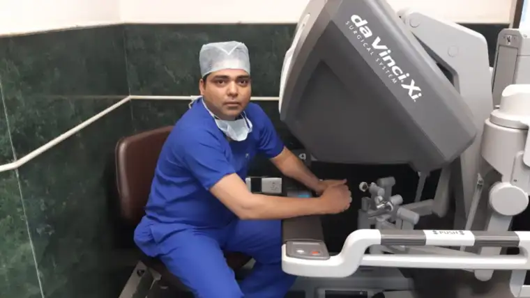
Dr. Arif Akhtar is a urologist practising in Gurugram with a focus that includes endourology and minimally invasive stone surgery — RIRS, PCNL and mini-PCNL — alongside his broader uro-oncology, robotic urology and renal transplant practice. Exact current qualifications, fellowship details, hospital affiliation and years of practice should be published exactly as verified by the doctor and kept consistent with the About page.
Your consultation includes a review of your imaging and reports, relevant blood/urine tests, and a discussion of which procedure suits your specific stone, along with any pre-procedure instructions.
Most kidney stone procedures are done under anaesthesia and, depending on the procedure and stone complexity, may be done as a short-stay or single-day admission. Your surgical team will explain the specific plan and expected hospital stay for your procedure in advance.
Recovery time varies with the procedure performed. A temporary DJ stent is often placed after RIRS or PCNL to help the kidney drain while it heals, and is removed at a follow-up visit. Because kidney stones can recur, follow-up also includes guidance on hydration, diet and, where relevant, further metabolic evaluation to reduce the chance of future stones.
Get clear answers to common questions about kidney stone treatment, RIRS, PCNL, recovery, recurrence prevention, and treatment costs in Gurugram.
Many smaller stones pass on their own with fluids and pain management, or can be treated with non-invasive shockwave lithotripsy (ESWL). Whether surgery is needed depends on stone size, location and symptoms, assessed individually.
RIRS uses a flexible scope passed through the natural urinary passage with no external incision, and suits many small-to-moderate kidney stones. PCNL accesses the kidney through a small incision in the back and is generally used for larger or more complex stones. Your surgeon will recommend the option best suited to your stone.
RIRS and PCNL are performed under anaesthesia, so you don't feel the procedure itself. Some discomfort during recovery is normal and is managed with medication; your care team will guide you on what to expect for your specific procedure.
Kidney stones can recur, particularly without changes to fluid intake, diet or an underlying metabolic cause. Follow-up and, where appropriate, stone-composition analysis help guide a personalised prevention plan.
Cost depends on the procedure recommended (ESWL, RIRS, PCNL or mini-PCNL), hospital stay and the treating hospital. Speak with the clinic directly for a personalised estimate after evaluation.