
Dr. Arif Akhtar
Senior Urologist
Hello! 👋 Welcome to Dr. Arif Akhtar. He is a Gurugram-based urologist specializing in robotic urology, uro-oncology, and kidney transplantation.
Just now
If a stent, dilation or endoscopic procedure has already been tried for a narrowed ureter and the problem hasn't resolved, that's a common pattern — not a sign the wrong treatment was chosen at the time. It's also exactly why ureteric reconstruction exists as a distinct, more definitive category of surgery. Dr. Arif Akhtar performs ureteric reconstruction in Gurugram, selecting from several established techniques based on precisely where and how long your stricture is.
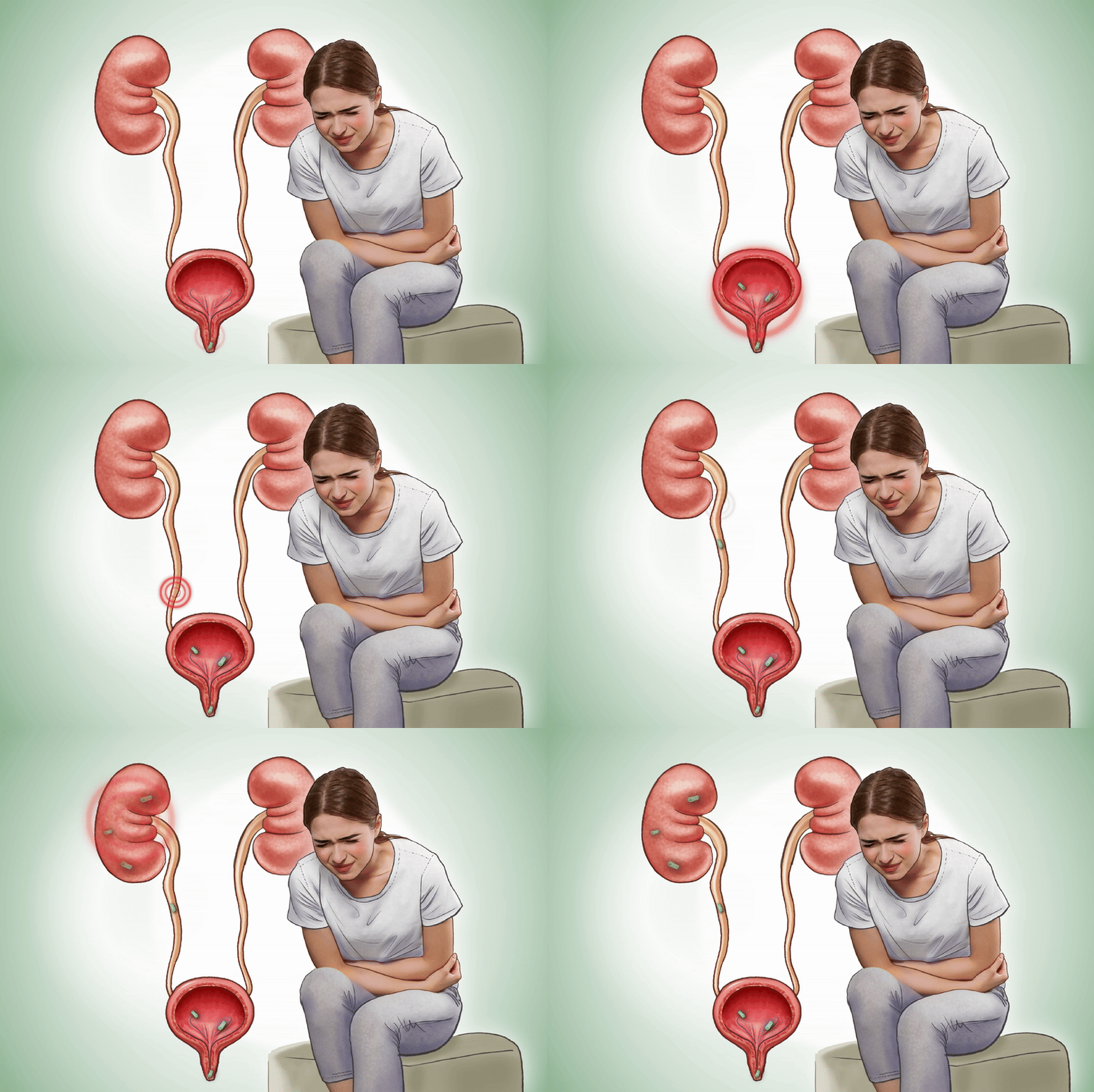
The ureter is the tube that carries urine from each kidney down to the bladder. A stricture is a narrowed or scarred segment of this tube that restricts urine flow. Left untreated, urine can back up toward the kidney (hydronephrosis), and over time this back-pressure can permanently damage kidney function — which is why a stricture, even one causing minimal symptoms, generally shouldn't be ignored.
Schedule a Clinical EvaluationUreteral strictures develop from scarring due to prior medical procedures, trauma, or underlying infections. Common contributing causes include:
Surgical & Stone Procedures: Scarring after prior kidney stone surgery or other urological interventions.
Surgical & Physical Injury: Injury during pelvic or abdominal surgery for unrelated conditions, or direct abdominal trauma.
Radiation Exposure: Radiation treatment targeted at nearby pelvic or abdominal cancers.
Infections: Infections including urinary tuberculosis, which remains a locally relevant cause in India.
Flank Pain & UTIs: Common symptoms include persistent flank pain, recurrent urinary infections, or a sense of fullness in the flank.
Asymptomatic Presentation: Many strictures cause no symptoms and are found incidentally after kidney function has already begun to decline.
Comprehensive Clinical & Imaging Assessment Methods
Accurate imaging and functional studies pinpoint the stricture's exact location, length, and effect on kidney health, forming the bedrock of a successful reconstructive surgical strategy.
A retrograde pyelogram — an X-ray taken while contrast dye is introduced into the ureter — or a CT urogram maps the exact location and length of the stricture, which is the single most important information for planning the right reconstructive technique.
A nuclear renal scan assesses how well the affected kidney is still functioning and draining — vital both for surgical planning and understanding whether the kidney is likely to recover function once the blockage is corrected.
Dilation and endoscopic incision offer a quick, minimally invasive option, but evidence consistently shows higher long-term recurrence rates because they do not remove the scar tissue. Surgical reconstruction is the ideal next step when endoscopic treatment fails, when strictures are long, or to avoid reliance on temporary stenting.
The narrowed segment is surgically removed and the two healthy ends of the ureter are rejoined directly. This is the most straightforward option, perfectly suited to short strictures where the ends can be reconnected without tension.
For strictures located near the bladder, the ureter is reimplanted directly into the bladder wall. A psoas hitch — fixing the bladder upward toward a nearby pelvic muscle — is frequently added to bridge larger gaps without putting tension on the new connection.
For longer strictures that a psoas hitch alone cannot bridge, a flap of bladder tissue is shaped into a functional tube to replace the missing segment of ureter, effectively extending the bladder upward to meet the healthy upper ureter.
For long or complex strictures located higher up, a tissue graft harvested from the inner lining of the cheek (buccal mucosa) is used to patch and widen the narrowed segment. This advanced technique effectively reconstructs strictures that were previously difficult to preserve.
Reconstruction can be performed via open surgery, laparoscopically, or with robotic assistance depending on patient anatomy and stricture characteristics. Robotic assistance is particularly beneficial for the delicate, fine suturing these precise reconstructions demand.
Dr. Arif Akhtar brings dedicated expertise to complex reconstructive urology, focusing on organ preservation and long-term functional recovery. By combining modern diagnostic mapping with advanced open, laparoscopic, and robotic techniques, he delivers individualized reconstructive solutions tailored to restore clear urinary drainage and protect vital kidney function.
Your journey starts with dynamic imaging (CT Urogram or Retrograde Pyelogram) and renal function scans to evaluate stricture length, location, and the functional capacity of the affected kidney.
Based on your imaging, the appropriate surgical technique — ranging from primary repair to tissue grafting or bladder flap reconstruction — is performed via open, laparoscopic, or robotic approaches.
A temporary internal ureteral stent supports healing over several weeks. Follow-up imaging confirms smooth urinary drainage and kidney recovery before normal daily activities are fully resumed.
Clear answers regarding ureteral strictures, symptoms, diagnostic imaging, reconstructive options, and long-term kidney protection in Gurugram.
Yes. Left untreated, a stricture causes urine to back up into the kidney (hydronephrosis). Over time, this chronic pressure damages functional kidney tissue, which is why strictures require clinical evaluation even when they cause no pain.
Endoscopic dilation or incision widens strictures temporarily but carries high recurrence rates because scar tissue remains. Reconstruction removes or patches the diseased segment, providing a durable, definitive solution without long-term stent dependence.
The choice depends on the exact location and length of the stricture mapped via CT urogram or retrograde pyelogram, as well as the overall functional health of the affected kidney.
For long or complex upper strictures, a small graft taken from the inner cheek lining is used to patch and widen the narrowed ureter. The cheek site heals rapidly with minimal discomfort, providing optimal tissue for durable repair.
Yes. Robotic-assisted reconstruction offers high magnification and precise articulation needed for delicate microsuturing, resulting in smaller surgical incisions, reduced postoperative discomfort, and faster overall recovery.
Yes, a temporary internal stent (JJ stent) is placed inside the ureter to support smooth healing and prevent obstruction. It is typically removed during a brief clinic procedure a few weeks after surgery.